Products

UV and Light

The most important source of light and subsequent energy on earth is the sun. The sun emits energy that can be absorbed by organisms and molecules where it facilitates countless reactions, including photosynthesis, ozone production, vitamin D, and weather phenomena.

Ultraviolet radiation (UVR)

Sunburn and skin cancer are known to be correlated to the dose of UV radiation our skin encounters. UV radiation is emitted by the sun, and is a region in the electromagnetic spectrum between 400-200nm, that can be broken down into three categories: UVA which is between 400-320nm, UVB which is between 320-280nm, and UVC between 280-200nm. As the various types of UV radiation are at different wavelengths, they react differently with the atmosphere. Almost all of the UVC radiation, some of UVB and UVA is absorbed before it reaches the Earth’s surface. The dose of UVA, UVB and UVC changes as UV passes through the atmosphere mainly due to differences in rates of reflection, refraction, scattering and absorption. The change can be emulated on a UV index (Figure 1). A local UV index can be found in a variety of places reporting on meteorological data and forecasts. UVI depicts how much UV radiation is likely to be reaching the Earth’s surface and therefore which precautions should be taken to minimise human risk.

Figure 1. UV index

Figure 1. UV index

A UV index value of zero corresponds to no UVR reaching the Earth’s surface, which occurs at night time, and an index of 10 refers to roughly midday sun, when there is no cloud cover. UVR indices record well into the teens when the ozone layer reads low values. The index numbers are measured in watts/meters2, and the measurements reflect different wavelengths to be more harmful than others.

Note that the above precautions are provided for adults with moderately fair to dark skin tone, and that skin damage caused by sun exposure is accumulative over one’s lifetime.

Ozone

Figure 2. Production of ozone in the atmosphere.

Figure 2. Production of ozone in the atmosphere.

As light enters the Earth’s atmosphere, it comes into contact with molecules in the stratosphere. The stratosphere is situated about 10km above sea level and continues up until 50km above sea level. It contains the ozone layer, which absorbs UV radiation. The ozone layer is composed of ozone, a molecule containing 3 oxygen atoms. Oxygen molecules are good absorbers of UVC radiation, using this energy the oxygen bonds break, forming two separate oxygen atoms. These atoms can rejoin, recreating the oxygen molecule, or binding to an oxygen molecule ultimately to form an ozone molecule. Ozone molecules are good absorbers of UVB, and to a lesser degree, UVA. Ozone molecules absorb the energy provided by UVB and UVA to undergo dissociation, releasing oxygen atoms that can combine to form oxygen molecules. These two reactions are in equilibrium, where neither really dominates. Ozone is an important molecule in reducing the amount of harmful UV radiation that passes through our atmosphere, but at low altitudes, such as on the surface of the Earth, it becomes a somewhat toxic and unstable molecule.

The amount of UV light that reaches the Earth’s surface varies and is dependent on a number of factors:

  • Latitude: Near the equator, where the sun is directly overhead, the distance from the sun to the ground is the shortest, and the UV has to pass through the least amount of radiation. Therefore, on the equator, the UV intensity is the highest. Likewise, on the poles, the sun appears low in the sky, meaning the light has to pass though more radiation-absorbing atmosphere to reach the surface.
  • Elevation: At higher altitudes there is less atmosphere to absorb UV radiation, therefore individuals are exposed to a greater amount of UVR.
  • Proximity to an industrial area: photochemical smog produced in industrial processes contains artificially produced ozone. The ozone produced can absorb more UV radiation, but also poses significant health risks if it is low lying.
  • Weather conditions: cloud cover can reduce the UV levels on the surface, but often incompletely.
  • Reflection: some surfaces are able to reflect UV radiation, meaning there is a greater chance of getting sun burnt, even in shady places. Snow, sand, grass and water are good reflecting surfaces.
  • Time of day/year: during the middle of the day, the sun is at its highest position in the sky, resulting in the least distance for radiation to travel, and more exposure to UV light. The summer months also provide the most intense UV radiation, as the sun’s angle (zenith angle) is reduced.
  • Ozone layer: the depth of the ozone layer plays a vital role in reducing exposure to UV light. The ozone layer has been found to be thinning in the past few decades. This is presumably due to the release of ozone degrading chemicals by industry.

Ozone depletion

In the past few decades, it has been observed that the ozone layer is thinning over certain regions of the globe, particularly over Antarctica. This has been attributed to chemicals that have been released by industry since the 1930’s, particularly gaseous chlorine and bromine containing substances, such as chlorofluorocarbons (CFC’s) and bromofluorocarbons (BFC’s).

Ozone can be broken down by natural and manmade sources. Atomic chlorine and bromine are two significant contributors, and can be released into the atmosphere by large volcanic eruptions and are also present in chemicals, especially those that where used by industry decades ago, such as CFC’s and BFC’s. CFC’s have been produced as a substitute for toxic and flammable chemicals used in industry, such as ammonia. They’re non-toxic, non-flammable, and their very stable nature made them perfect for propellants in aerosol cans and refrigerants. Their use have grown exponentially (in 1988 it was estimated that over one billion kilograms were being produced annually worldwide) until the discovery that CFS had the capacity of thinning the ozone layer. At this stage world leaders acted by signing the Montreal Protocol, which stipulated that consuming and producing chemicals capable of breaking down ozone be phased out by 2000.

CFC and other similar chemicals are so stable that they’re not destroyed in the troposphere, as they slowly rise towards the stratosphere. These stratospheric molecules are found to be able to break ozone down into an oxygen atom and molecule. This usually occurs at a slow rate, reducing the overall thickness of the ozone layer by about 4% per decade, but in the atmosphere above Antarctica, especially in spring, thinning occurs at an alarming rate. During its winter, Antarctica becomes cold enough (-80°C) for stratospheric clouds (termed Stratospheric Clouds Mother of Pearl, PSCs) to form in the ozone layer. On the surface of these clouds, chlorine and bromine are transformed into an active state and when the sun gains intensity in the spring, clouds disappear freeing chlorine and bromine to rapidly destroy ozone. As temperatures further continue to warm, wind vortexes holding the particles above Antarctica break up, mostly allowing ozone rich air to flow into ‘the hole’. The chlorine and other molecules capable of breaking ozone down can destroy up to a thousand molecules of ozone before it is converted into an inactive form, such as hydrochloric acid.

There are many complex reactions that destroy ozone, the simplest being: a free chlorine atom reacts with an ozone molecule, forming a chlorine and oxygen complex, ClO and an oxygen molecule, O2. The ClO then reacts with a free oxygen atom forming an oxygen molecule and a free chlorine atom. In this manner a circular process is initiated.

Although the Montreal Protocol, first signed in late 1988, will help assist to reduce the effects of destructive radicals in the atmosphere, the effects will not be instantaneous. As it takes years for the CFC’s and other molecules to reach the stratosphere, the reduction in emissions will have minimal effect in the stratosphere (the largest dimension of the hole over Antarctica was measured in 2006, being over three times the size of Australia). It is anticipated that it will take a decade before confirmation of recovery of the ozone can be given.

With the thinning of the ozone layer, less UVB radiation is absorbed resulting in more harmful radiation passing to the Earth’s surface. This would result in increased incidence of skin cancer, cataracts, and other UV related diseases.

Definitions and focus

Figure 3. A typical wavelength.

Figure 3. A typical wavelength.


Figure 4. Electromagnetic spectrum.

Figure 4. Electromagnetic spectrum.

The energy in the light emitted by the sun and all other sources, travels as mass-less, charge-less particles, photons. Photons can travel as both particles and waves, where each beam of light has its own wavelength (measured peak to peak: Figure 3), frequency (number of wave cycles passing a point per second, Hertz) and energy.

The wavelengths of visible light range from 400nm to 700nm. Once the wavelength drops outside of these values a different form of electromagnetic radiation transpires; these different forms can be found on an electromagnetic spectrum (Figure 4). As evident on Figure 4, the two main sources of electromagnetic radiation emitted by the sun (visible light and ultraviolet radiation – UVR) only occupy a small section of the spectrum.

Environment

The heat supplied by the sun has provided us with a suitable environment to survive. Once radiating energy is passed through the atmosphere, the photons collide with matter, transferring energy. This causes heat, wind, rain, clouds and other weather phenomena. Wind is a result of the varying temperatures of the air above land and water. The hot air rises, causing a flow of cooler air to occupy its previous place, and eventually cool air, high in the atmosphere, drops down to sea level, completing the cycle. When ocean water is heated, water particles evaporate into the air, where they culminate to form clouds. These clouds pushed over land by winds experience lower temperatures, causing condensation into droplets and precipitation. Differences in atmospheric moisture, pressure and temperature result in dramatic weather conditions as hurricanes, cyclones and tornadoes.

When the sun’s rays are shone on an organism, it can have harmful consequences. Different types of electromagnetic radiation have various effects on tissue and DNA. The ultraviolet region of the electromagnetic spectrum can cause serious damage to organic tissues, including human tissue. Humans are protected against ultraviolet radiation by various natural defense mechanisms. Ultraviolet radiation (UVR) can also cause damage to our eyes, both short and long term.

For instance the conjunctiva of the eye may show inflammatory reactions when exposed to intense UVR. Long-term eye (ophtalmis) exposure to sun may have deleterious effects on the retina, causing immediate, and sometimes permanent, visual loss. Long-term UV exposure contributes to cataract formation.

Photosynthesis

Figure 5. The process of photosynthesis.

Figure 5. The process of photosynthesis.

Photosynthesis is a process carried out by photo-autotrophs.Put simply, photosynthesis is water and carbon dioxide, in the presence of light, forming oxygen and glucose.

The oxygen produced is released out into the atmosphere through stomata while the carbohydrate, glucose, can be modified to produce various other carbohydrates, such as cellulose for plant walls, or starch, a glucose storage molecule.

The carbon dioxide is acquired through stomata, small gaps in plant leaves that are surrounded by two guard cells. These guard cells are able to open and close depending on the environmental conditions. For instance, if conditions are extremely dry, guard cells would swell to close the gaps, in order to minimize water loss. These gaps allow carbon dioxide and water vapor to enter the plant, and oxygen and water vapor to leave. In turn, water is acquired through the plant roots. Moisture obtained from the soil may travel through the roots up the plant to the leaves via the xylem (vascular tissue). The xylem is a tissue in vascular plants that carries water and minerals from the roots, to all other sections of a plant.

Figure 6. Diagram of a chloroplast.

Figure 6. Diagram of a chloroplast.

The energy within the sunlight is captured by chlorophyll, a pigment found in chloroplasts. Chlorophyll is of biological relevance due to its chemical structure, and subsequent absorption spectra. Also present in chloroplasts and involved in photosynthesis are grana, which are stacks of thylakoid discs, and the stroma, which is a dense fluid surrounding the grana (see Figure 6).

Photosynthesis is separated into two phases: the light dependent and independent stages. The light dependent stage involves the absorption of a photon and its conversion to an electron, a series of reactions follows, which conclude in an oxygen molecule being released into the atmosphere. The light independent reaction involves the absorption of carbon dioxide by an enzyme, RuBisCO, and results in the production of carbohydrates.

Light dependent reaction

Figure 7. Diagram of a photosystem.

Figure 7. Diagram of a photosystem.

The light dependent reaction can occur by two mechanisms: cyclic photophosphorylation and non-cyclic photophosphorylation. Non-cyclic photophosphorylation is the more common of the two in plants, produces ATP and NADPH and involves the use of two photosystems, photosystem I and II. Cyclic photophosphorylation occurs in plants when there is insufficient NADP+ to produce NADPH, involves photosystem I and only produces ATP.

A photosystem is an arrangement of pigments, such as chlorophyll, packed into a thylakoid membrane. The energy containing packets in light (photons) are absorbed by accessory pigments, passed to the primary pigment, and then collected where they initiate the production of energy in the form of ATP and NADPH. The primary pigment associated with photosystem I is P700, while P680 is associated with photosystem II. These numbers 700 and 680, refer to the wavelengths of light that the respective pigments can absorb.

Figure 8. Non-Cyclic photophosphorylation reaction.

Figure 8. Non-Cyclic photophosphorylation reaction.

Non-cyclic photophosphorylation (Figure 8) begins with photosystem 2. The photons present in light are absorbed by P680, where they excite an electron. This electron is then passed to a primary electron acceptor molecule through a process termed photoinduced charge separation. The positive charge left on the P680 molecule is alleviated by the extraction of electrons from water molecules, causing the release of atomic oxygen. The electron held by the primary electron acceptor is passed to pheophyton then down the electron transport chain. This creates a chemiosmotic gradient which leads to ATP being formed by ATP synthase in the light independent reaction. Once passed through the electron transport chain, the electron enters photosystem 1 where it is re-excited and begins its path down another electron chain. The electron is again passed through a series of reactions, eventually being added to NADP+ reductase to form NADPH.

Figure 9. Cyclic photophosphorylation reaction.

Figure 9. Cyclic photophosphorylation reaction.

Cyclic photophosphorylation only involves photosystem 1. P700 absorbs photons until an electron is raised to its excited state. The electron then passed undergoes photoinduced charge separation, before being passed down an electron transport chain, producing a chemiosmotic potential across the membrane. This ion gradient stimulates the production of ATP from ATP synthase. Once the electron reaches the end of the electron chain, it is passed back to the photosystem by electron acceptor molecules (see Figure 9). Cyclic photophosphorylation also occurs in certain photosynthetic bacteria.

Light independent reaction

Figure 10. Light independent reaction.

Figure 10. Light independent reaction.

Light independent reactions are also referred to as carbon fixation. Ribulose biophosphate (RuBP), with assistance from the enzyme RuBisCo, is combined with a carbon dioxide molecule. The end product is two, three-carbon, 3-phosphoglycerate (PGA), molecules. The ATP and NADPH produced in the light dependent reaction are used to reduce PGA to 3-phosphoglyceraldehyde (PGAL). Additionally, 5 of every 6 molecules of PGAL produced are reused in the production of RuBP, while the remaining molecule is used in the formation of carbohydrates, such as starch, cellulose and glucose (Figure 10).

References

  • Cartage.org, (2007). Photosynthesis [Online]. Available from: http://www.cartage.org.lb/en/themes/sciences/BotanicalSciences/Photosynthesis/Photosynthesis/Photosynthesis.htm [Accessed on 29/04/2008].
  • de Gruijl, F R, Longstreth, J, Norval, M, Cullen, A P, Slaper, H, Kripke, M L, Takizawa, Y, (2003). Health effects from stratospheric ozone depletion and interactions with climate change, Photochemistry and Photobiology. Vol 2, pp 16-28.
  • Helpsavetheclimate.com, (2007). Photosynthesis [Online]. Available from: http://www.helpsavetheclimate.com/photosynthesis.html [Accessed on 29/04/2008].
  • Slaper, H, Velders, G J, Daniel, J S, de Gruijl, F R, van der Leun, J C, (1996) Estimates of ozone depletion and skin cancer incidence to examine the Vienna Convention achievements, Nature. Vol 384(6606). pp 256-258.
  • Solcomhouse.com, (2006). Ozone [Online]. Available from: http://www.solcomhouse.com/ozone.htm [Accessed on 29/04/2008].
Rare Skin Conditions

Congenital Erythropoietic Porphyria (CEP)

Snapshot Other common terms: CEP, Gunther’s disease, Uroporphyrinogen III synthase deficiency, UROS deficiency, Congenital porphyria, Congenital hematoporphyria, Erythropoietic uroporphyria ICD-10 classification: E80.0 Prevalence: Extremely rare; less than 200 cases reported. Usually manifests during infancy or early childhood. Causes: Genetic mutation in the UROS gene leading to reduced enzyme function. Symptoms: Symptoms may include: blistering, scarring, necrosis or excessive hair growth on light-exposed skin; disfiguration of the ears and nose; loss of fingers; anaemia; red-stained teeth; pink/red coloured urine and enlarged spleen. Treatments/cures: Treatment with blood transfusions, splenectomy, oral sorbents, beta-carotene…
Read More…

Actinic Prurigo (AP)

Snapshot Other common terms: AP, Hutchinson prurigo ICD-10 classification: Not defined, L55-59 Prevalence: Unknown. More common in Latin and Indigenous Americans Causes: Not well understood. Suggested that an immune-mediated response to UV light is responsible. Symptoms: Extremely itchy skin rash, red and inflamed bumps (papules), thickened patches (plaques) and/or lumps (nodules) following exposure of skin to sunlight. Treatments/cures: In some cases, actinic prurigo may resolve itself. Topical steroids, emollients, phototherapy, thalidomine and oral immunosuppressants. Differential diagnosis: Polymorphous light eruption, prurigo nodularis, lupus Actinic Prurigo (AP) is a rare chronic, idiopathic…
Read More…

Erythropoietic Protoporphyria (EPP)

For more on Atopic’s clinical program for EPP, click here. Snapshot Other common terms: EPP, protoporphyria, erythropoietic porphyria ICD-10 classification: E80.0 Prevalence: Rare; between 1:58,000-200,000. Estimates of between 5000-10,000 globally Causes: Inherited disease; defective enzyme causes inability to properly produce haem (heme). Symptoms: Phototoxicity: swelling, burning, itching and redness of the skin, occurring during or after exposure to sunlight, including light passing through windows. Liver toxicity in 5% of cases. Microcytic anaemia can occur. Treatments/cures: None proven fully effective to date. Phototoxicity can be avoided by complete avoidance of sunlight…
Read More…

Hydroa Vacciniforme (HV)

Snapshot Other common terms: HV, Bazin’s Hydroa Vacciniforme ICD-10 classification: L56.8 Prevalence: Rare. Prevalence data is scarce, reported as 0.34:100,000 in Scotland. Mainly presents in children aged 3-15. Causes: Exact cause unknown; there is correlation between diease symptoms and exposure of skin to UV (particularly UVA) radiation. Symptoms: Eruption of fluid-filled blisters on the skin following exposure to UV radiation. Treatments/cures: No known cure, HV commonly resolves in late adolescence. Phototherapy, fish oil and various drugs may reduce symptoms. Differential diagnosis: Porphyrias (erythropoietic protoporphyria, congenital erythropoietic protoporphyria), Polymorphous Light Eruption,…
Read More…

Porphyria Cutanea Tarda (PCT)

Snapshot Other common terms: PCT ICD-10 classification: E80.1 Prevalence: Uncommon; 1:25,000 Causes: Inherited disease; defective enzyme causes inability to properly produce haem (heme). Symptoms: Skin photosensitivity causing extremely fragile skin and changes in pigmentation (melanin). Discoloured urine. Treatments/cures: Cannot be cured. Avoidance of sunlight and certain artificial lights. Differential diagnosis: Erythropoietic protoporphyria, polymorphous light eruption Porphyria cutanea tarda (PCT) is the most frequently seen disease of a group of disorders (the Porphyrias) that can be acquired or inherited. It is caused by low levels of an enzyme (uroporphyrinogen decarboxylase or…
Read More..

Solar urticaria (SU)

Snapshot Other common terms: SU ICD-10 classification: L56.3 Prevalence: Very rare; 3.1:100,000 individuals globally. Solar urticaria is more likely to affect women Causes: Exposure of skin to light. Exact allergen is unknown. Symptoms: Systemic: anaphylaxis, breathing difficulty, nausea and headaches. Immediate localised reactions on skin: characteristic ‘wheal’ formation, erupting flares on exposed skin sites and to swelling of soft tissues. Treatments/cures: No known cure. Anti-histamines and topical steroids may be useful in some cases. Immunosuppressants and plasmaphoresis in extreme cases. Differential diagnosis: Polymorphous light eruption, drug induced photosensitivity, other allergies/physical…
Read More

Urticaria Pigmentosa (UP)

Snapshot Other common terms: UP ICD-10 classification: Q82.2 Prevalence: Rare, exact prevalence is unknown but it is more common in Caucasians Causes: Excess of inflammatory mast cells due to an unknown cause; mast cells trigger histamine in the affected area. Environmental factors may trigger symptoms. Symptoms: Swelling, itchiness and a rash on the skin. May present as brown patches, hives, welts, rashes, blisters or facial flushing. Diarrhea, low blood pressure and an increased heart rate may present in certain cases. Treatments/cures: Avoidance of causes. Anti-histamines, mast cell stabilizers and topical…
Read More…

Xeroderma Pigmentosum (XP)

Snapshot Other common terms: XP ICD-10 classification: Q82.1 Prevalence: 1:250,000 globally except in Japan where incidence is 1:40,000 Causes: Inherited. Autosomal recessive disease. Symptoms: XP causes a defect in DNA repair, making affected individuals hypersensitive to UV light and causing an extreme susceptibility to skin cancers. Ocular and neurological issues are likely. Treatments/cures: No known cure. Treatment is limited to sun avoidance, and immediately treat skin cancers. Xeroderma Pigmentosum (XP) is a rare, hereditary disease where patients experience skin hypersensitivity to ultraviolet (UV) light. It is characterized by dry skin,…
Read More…

Melanin in Biology

Melanin is a complex polymer synthesized by living organisms. A polymer is a large chemical compound often made up of hundreds of smaller chemical parts, monomers. A variety of melanins can be found through biology, most commonly being composed of derivatives of acetylene, a black monomer.

Human Melanin

Melanins are found in a wide variety of organs in humans. They have been found in a few regions of the brain, where they are termed neuromelanins. Their exact function is unknown, but it has been suggested that they could be a byproduct of the synthesis of a neurotransmitter. Neuromelanin is probably the only source of these specific neurotransmitters with a variety of neurodegenerative compounds coupled with a reduction in neuromelanin. In the inner ear melanin protects against hearing damage, this is presumed to be because it has been found that it is the best sound absorber known. In the eye it protects the iris against UV damage, similar to the way it protects the skin. It is has also been discovered in the birth canal and adrenal gland. All of these melanins have different structures, they might be slight alterations of each other, or distant relatives.

Melanin in the skin

Human skin and hair contain both the reddish pigment pheomelanin and brownish eumelanin, where eumelanin is the most abundant in the human body. Pheomelanin is more present in females, leaving females with a more reddish skin.

Where reference is made to melanin in the text, eumelanin is implied. The precise function of melanin is the subject of much debate; its exact role is still unclear. In the dermis (skin), the pigment is a primary determinant of both skin and hair colour. Melanin is produced by melanocytes, special skin cells located in the basal layer of the epidermis (Figure 1). The process of activating pigmentation in the skin is termed melanogenesis, and is initiated after cellular damage following exposure to UV radiation or the sun. Visibly, increased melanin in the skin results in a darker skin complexion, and better protection.

Figure 1. Melanosomes leaving the melanocyte as a result of sunburn in the upper epidermis.

Figure 1. Melanosomes leaving the melanocyte as a result of sunburn in the upper epidermis.

Both melanins are synthesized in the basal layer, which is located at the bottom of the epidermis (top layer of the skin), above the dermis (see Figure 1). Special cells, melanocytes, produce melanin containing packets, called melanosomes. These melanosomes are spread throughout the epidermis by tentacle-like projections called dendrites. The dendrites, which are connected to the melanocytes, transfer the melanosomes to separate keratinocytes (skin cells). Once the melanosomes reach the end of the projections they are squeezed out, into the keratinocytes. The melanin containing packets spread out above the nucleus, where they stay, protecting the DNA inside the organelle from harmful UV radiation. The skin cells eventually rise to the top of the epidermis where they die and are desquamated (shed away).

All humans appear to almost have the same number of melanocytes and keratinocytes (at a ratio of 1:36). The size and number of melanosomes, however, can vary dramatically between individuals. People with fair skin tend to produce small melanocytes, containing little melanin, which is portrayed as a fine brown dust covering the nucleus of keratinocytes. Dark skin individuals produce larger melanin containing packets that are seen as dark patches covering the nucleus. Both of these forms of melanosomes protect the DNA in two ways, by scattering incoming ultraviolet radiation and by absorbing it. Because of this protective ability that melanin posses, it is called a photoprotectant, that is a substance that can protect an organism from damage caused by the absorption of photons from a radiating source, particularly the sun. This degradation is called photodegradation.

The more incoming UV radiation that is randomly scattered, the less the chance of DNA’s exposure to it. The amount and direction of the scattering is dependent on both the wavelength of the incident ray, and the size of the particle that it is striking.

Research has shown that the smaller a particle size, the greater the evenness of scattering. It has been revealed that even though some radiation passes through the melanin, if the layer is thick enough, it will eventually be dispersed or absorbed elsewhere.

Figure 2. Scattering caused by different sized particles when subject to the same wavelength of light.

Figure 2. Scattering caused by different sized particles when subject to the same wavelength of light.

The sizes of human melanosomes range from around 228nm to 684nm. It is therefore desired, on a scattering basis, to have melanosomes closer to 230nm rather than 690nm.

On the other hand, light absorption is favored by a larger surface area, which requires larger melanosomes. Once the energy is absorbed by the melanin, it raises electrons to excited states and, in turn, passes the energy to the cell it is situated in. The cell is then assumed to use this energy to regulate its conditions and drive chemical reactions, a role similar to that of chlorophyll in photosynthesis.

As a photoprotectant, melanin can be artificially produced and incorporated into products and substances that seek to offer protection against UV radiation. In other domains, similar types of melanin have also been used in conjunction with plastics, plastic films, and optical lenses.

Organisms

In the same way that melanin provides skin and hair colour, it also provides the colour of birds’ plumage, amphibians’ and reptiles’ skin or scales, and other animals’ fur. Pigmentation in animals provides bright colours that attract mates in the animal kingdom and is used for camouflage, as illustrated by the cuttlefish, which releases an ink composed of melanin particles, to distract and confuse its predators. It is thought to also be involved in the defense of fungi and bacteria against ionizing UV radiation, chemical strain and biochemical strain.

Additionally, in pathogenic microbes, melanin is used to protect the microbe from immune responses by its host. Also, in some organisms, melanin is involved in a self-defense mechanism, it encapsulates invading pathogens and is accompanied with the production of radical byproducts, it aids in the killing of pathogens.

Recently, due to the presence of melanin, fungi have shown to survive under harsh radioactive conditions. It is thought that melanin is converting ionizing radiation into a useable energy source, which promotes growth. This was first noticed after the melt down at the Chernobyl nuclear power plant in Ukraine. A sample taken of a black fungus found inside a reactor was found to contain melanin, triggering the speculation that the polymer melanin was harnessing the energy from the radioactive rays into a usable energy.

Melanin structure

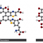
The chemical structure of the different forms of melanin have proven difficult to analyze, due to the properties of the molecule. They are found to be insoluble, amorphous, and cannot be studied as a solution or in crystal form. To overcome the frustrating properties, a partial degradation process has been used to study the separate components of each melanin structure. Two of the main melanins in the human body are eumelanin and pheomelanin (Figure 3).

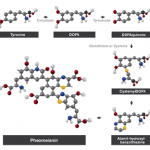
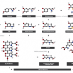
The pathway to synthesize melanins is also not fully understood. It is known that tyrosine is the starting reactant, and tyrosinase the enzyme that catalyses the reaction (Figures 4 and 5).

References

  • Chedekel, Zeise, Fitzpatrick (1994) ‘Melanin: Its role in human photoprotection’, Publish city: United Kingdom. John Murray.
  • Dadachova, E, Bryan, R A, Huang, X, Moadel, T, Schweitzer, A D, Aisen, P, Nosanchuk, D J, Casadevall, A. (2007). ‘Ionizing Radiation Changes the Electronic Properties of Melanin and Enhances the Growth of Melanized Fungi’ PLoS ONE Vol 2(5).
    Miyamoto, K, Baba, K, (1987) ‘Stereological Method for Unfolding Size-Shape Distribution of Spheroidal Organelles from Electron Micrographs’, Journal of Electron Microscopy, Vol 36(3) pp 90-97.
  • Nosanchuk, J D, Casadevall, A. (2006). Impact of Melanin on Microbial Virulence and Clinical Resistance to Antimicrobial Compounds, Antimicrobial Agents and Chemotherapy . Vol 50, pp 3519-3528.
  • pg.com (1997), Skin Pigmentation [Online]. No longer available online. Accessed on 29/04/2008.
Skin Layers

The skin (dermis, cutis, derma, integument, and cuticle) is the largest organ of the human body (approximately 20 square feet), weighing approximately 16% of bodyweight. Skin thickness varies and is on average 1mm thick; the thinnest on eyelids at 0.5mm and the thickest at 1.5mm on the palms and soles. Skin consists of multiple layers (stratified), epidermis, dermis and hypodermis (deepest layer).

Epidermis

Epidermis

Skin cross section showing the epidermis (not to scale)

The upper most outer layer of the skin, the epidermis, consists of squamous cells (flat and scale-like in shape) and underneath basal cells (round shaped). The process of keratinisation (formation of horny layer) occurs over the course of 4 to 6 weeks when keratinocytes (85% of the epidermal cells) migrate upwards through the skin to be desquamated (shedding of old skin) at the skin surface. Renewal of the upper layer is continuously initiated by the keratinocytes.

The mitotic layer (single line) is the area where the division of keratinocytes takes place, held together by desmosomes (macula adherens) resisting shearing of the epidermal layer. Mitosis (cell division) occurs in the ratio of 1:2, with each keratinocyte producing two identical new cells. One cell remains in place to enable it to divide again, as the other migrates to the differentiation layer, the upper and thickest layer of the epidermis (5-15 cells thick). After initial growth, a newly formed layer, the Malphigian layer, gives rise to the production of keratin, an insoluble sulphur-based fibrous protein. In the Malphigian layer, Langerhans cells play a role in the recognition of antigens and interaction with epidermal T-cells (lymphocytes), as an immunologic response.

In the deepest layer of the epidermis, called the stratum basale, Merkel cells are found, these cells are part of the cell-neuron complex: a close association with an afferent nerve ending, the sensory part of the epidermis.

Keratinisation

A cross section of skin highlighting keratinocytes, the epidermis and dermis (not to scale)

A cross section of skin highlighting keratinocytes, the epidermis and dermis (not to scale)

As the keratinocytes migrate upwards, they lose their content of the cell nucleus (anucleate) and become flat in structure. When reaching the outermost layer of the epidermis, the keratinocytes become corneocytes, with relatively little biological activity. This is the end-stage of the keratinocytes. The keratinocyte at this stage is filled with keratin and ceramides; these are lipids and fatty acids, enabling the dermis to retain moisture. The most external epidermal layer in contact with the external environment is the stratum corneum. The external corneocyte is distinguished in 2 layers, a compact and a sloughing layer. When the junction, corneodesmosomes, between these layers breaks down, desquamation (skin shedding) occurs.

Melanocytes and melanogenesis

A cross section of skin highlighting melanocytes, the epidermis and dermis (not to scale)

A cross section of skin highlighting melanocytes, the epidermis and dermis (not to scale)

The deeper layer of the epidermis contains the pigment producing (melanogenesis) cells, melanocytes. These produce the dark brown pigment in the epidermis, eumelanin. The melanocyte forms part of the epidermal unit in a ratio of 1:36 keratinocytes. Melanin production is initiated through the up regulation of the receptors on the melanocyte, called the melanocortin 1 receptor (MC1R). The melanocyte produces intracellular eumelanin through a tyrosinase mediated biochemical pathway. Once eumelanin is formed within the cell organelle melanosome, the eumelanin granules are transferred after dendrite formation by the melanocyte.

Melanin has a photoprotective function (shields against UV and light damage) to the skin. The second form of melanin is pheomelanin, reddish-yellow pigmentation. Pheomelanin is the major type of pigmentation in red hair and also predominates in the epidermis of skin types I and II (fair skinned individuals). Eumelanin, on the other hand, is present in large amounts in individuals with dark skin and hair (skin types III-VI). It is generally accepted that eumelanin provides greater photoprotection than pheomelanin.

Dermis

A cross section showing the dermis (not to scale)

A cross section showing the dermis (not to scale)

The dermis is the deeper skin layer consisting of two layers: the papillary and elastic, both containing collagen. Blood vessels (capillaries) and nerves are found in this layer.

The principle structural component of the dermis is the protein collagen, produced by fibroblasts dispersed throughout the dermis. A quantity of collagen molecules are bundled together throughout the dermis, making up for three-quarters of the dry weight of skin. Collagen is mainly responsible for dermal strength, representing connective tissue, the glue or cement holding together smooth muscles tissues found throughout the body. Collagen is, along with elastin, a pivotal component of bones, cartilage, tendons, the skin, lung tissue and blood vessels. Most of all, collagen is the main structure to provide firmness to body tissues, while elastin provides flexibility to these tissues.

As part of the dermal sensory system, nerves transmit and evoke pain, itch, and temperature from the dermis. Specialized nerve cells, Meissner’s and Vater-Pacini corpuscles, transmit the sensations of touch and pressure. Also present in the dermis are eccrine, apocrine, sebaceous glands and hair follicles.

Hypodermis

The hypodermis contains specialized cells and structures, and adnexae, such as hair follicles, each follicle being attached to the arector pili muscle. Oil, scent and sweat, sebaceous, apocrine and eccrine glands are all associated with the follicle. The hypodermis is characterized with a high density of lipocytes, adipose or fat cells. The adipose layer serves as the largest reservoir of fatty acids. There is gender difference in distribution and size of adipose cells (in females predominantly in the buttocks and thigh area; in males in the abdominal region). This hypodermis also contains sweat and eccrine glands.

Hair is regarded as “dead epidermal cells” that have evolved through continuous modified epidermal keratinization. Upon close examination, hair is characterized by the expression of specific keratin proteins that are intensely cross-linked by disulfide bonds. Hair stems from hair follicles, which are epidermal invaginations that project into the dermis or hypodermis. There are two types of hair: vellus – fine short, soft, fine, and pale hair; and terminal – thick hard, large, coarse, long and dark hair. The number of hairs on all primates is similar, but mostly seen is vellus on humans and terminal on other primates. Hair can be “mobilized” by arector pili muscles, a smooth muscle enabling hair to stand on end most likely for better insulation (the phenomenon of goose bumps/pimples). The hair follicle undergoes a cycle of active and resting phases during which a new hair is started and then falls out, respectively. Melanocytes are seen to be scattered throughout the hair shaft. The melanocytes render colour to the hair. With age, tyrosinase production decreases and the hair turns gray.

Eccrine sweat glands are simple coiled tubular glands located in the deep dermis or underlying hypodermis and are present throughout the body. They develop as invaginations of the epithelium of the epidermal ridge and grow into the dermis, and the deep aspect eventually develops into the glandular portion of the sweat gland. Eccrine sweat glands have two regions: a secretory region and a duct region. The secretory portion is comprised of simple coils of cuboidal epithelium containing two kinds of cells. Dark cells produce sialo-mucins, while clear cells produce water and electrolytes. Myoepithelial cells support and constrict the gland in response to cholinergic stimulation. Secretion is controlled by heat stress in most of the body but is under emotional control in palms and soles. The duct portion of the sweat gland is stratified cuboidal epithelium (2 layers), whose cells resorb ions (Na+, K+, Cl-) from the glandular secretion. The final product is hypotonic (99% water) containing salts, lactate and urea. Adults are able to produce between 0.5-1.0 litre/day. Apocrine sweat glands are simple tubular glands that empty into hair follicles in axillary and anogenital regions. The secretion is a mixture of proteins, carbohydrates, and ferric ions, odourless when secreted, but which is acted on by commensal bacteria. They begin to function at puberty; but their function remains unknown.

Function of the skin

Human skin has numerous functions, it is the major interface between the environment and the human organs and so it serves many specialised functions that facilitate survival. It regulates body temperature to protect against hyperthermia and hypothermia. Water loss is controlled to protect against dehydration and is involved in controlling a balance of body fluids, mineral and waste product loss.

Skin also protects from the invasion of noxious substances, UV light, heat and micro-organisms. Langerhans cells have been found to be involved in a number of reactions to protect against micro-organism invasion and pain. They have antigen-presenting capacity, force keratinocytes to secrete immune regulating cytokines and T-cells, and are involved in delaying hypersensitivity.

The skin is also the most extensive sensory organ of the body for detection of tactile, thermal and painful stimuli for the start of vitamin D production – which is pivotal for bone growth – and serves other important immunological functions. Wound healing of the skin is regulated by an intact immunological defense mechanism.

References

  • Marks, R (2004). ‘The Stratum Corneum Barrier: The Final Frontier’, The Journal of Nurtition, Vol 134(8). pp2017-2021.
  • Jablonski, N & Chaplin, G. (2000), ‘The Evolution of human Skin Coloration’ Journal of Human Evolution. Vol 39. pp 57-106.
  • Chedekel, M R, Zeise, L & Fitzpatrick, T B (1994). Melanin: Its Role in Human Photoprotection. Overland Park: Valdenmar Publishing Company.
  • Anderson, R R & Parrish, J A. (1981), ‘ The optics of human skin’, Journal of Investigative Dermatology. Vol 77(1). pp 13-19.